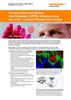
세포 생물학
라만 분광법은 비침습적, 비표지 분석 기법입니다. 유전자 조작이나 스테인 또는 항체 사용 없이 화학 정보를 추출합니다. 따라서 세포의 진정한 화학 정보를 확보할 수 있습니다.
세포 식별
다음과 같은 세포를 식별 및 구별하는 데 Renishaw 라만 시스템을 사용하십시오.
- 정상 세포 중 암 세포
- 분화된 세포 중 줄기 세포
- 세포 집단 중 다른 하위 상태(예: 줄기 및 전구 세포)
알려진 마커없이도 고유의 화학 프로필을 기반으로 세포를 식별할 수 있습니다. 항체와 결합하거나 유전자를 조작할 필요가 없습니다.
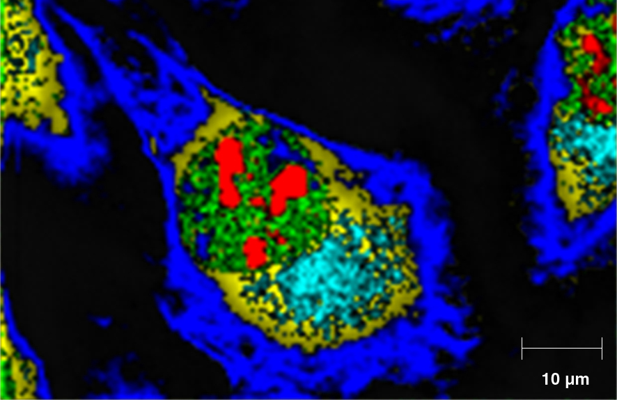
미세한 생물학적 정보 확인
우수한 공간 분해능의 칸포칼 라만 분석으로 검사할 수 있는 대상은 다음과 같습니다.
- 현장에서 개별 세포들의 세포 내 구조와 생체 분자
- 효모 세포의 화학적 성분
- 지질 대사를 정확히 이해하는 데 유용한 암 세포의 지질 함량
집단 내 개별 세포를 연구하고 세포 간 다양성을 판정할 수 있습니다. 예를 들어 건강한 세포와 비정상 세포의 지질 및 DNA 분포를 분석할 수 있습니다.
생체 세포 연구
Renishaw 라만 시스템에 세포 배양기를 장착할 수 있습니다. 이 챔버는 분석하는 동안 정상적인 생리학적 상태로 세포를 유지하기 위해 온도, CO2 농도 및 습도를 제어할 수 있습니다.
생체 세포 데이터는 종점 실험보다 모든 역동적 프로세스를 더 정확히 보여줍니다. 예를 들어, 해당 환경이나 약물의 변화에 대한 세포의 반응을 모니터링 할 수 있습니다. 이러한 반응은 모든 대사나 형태학적 변화 또는 세포 사멸(아포토시스)로서 세포를 나타낼 수 있습니다. 로만 분광기를 통해 이 모든 현상을 확인할 수 있습니다.
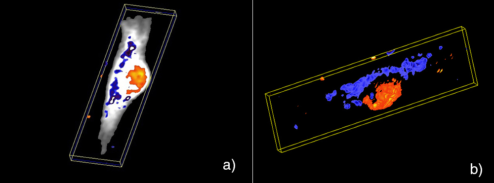
3차원으로 세포 내 변화 확인
화학 정보를 수집하고 샘플의 3D 보기를 생성하여 세포에 의한 물질 흡수를 확인하는 데 사용해보십시오. 세포와 세포 기관의 볼륨도 확인할 수 있습니다.

다운로드: 생명 과학(세포)
-
 Application note: Classification of brain glioma tumours using the Renishaw Biological Analyser [en]
Demonstrating discrimination between diseased and healthy brain tissue using the Renishaw RA816 Biological Analyser.
-
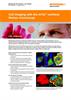
 Application note: Cell imaging with the inVia confocal Raman microscope [en]
With Renishaw’s inVia confocal Raman microscope you can identify and characterise samples to provide chemical, spatial and structural information on multiple types of molecule, without labelling. It provides rich, detailed, chemical images and highly specific data at high spatial resolution, making it ideal for studying cells.
-
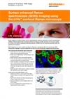 Application note: Surface enhanced Raman spectroscopy (SERS) imaging using the inVia confocal Raman microscope [en]
Raman imaging is a powerful research tool for understanding the molecular composition, structure and distribution of different chemical species. Nano silver/gold colloids and roughened metallic substrates can be used to amplify the intensity of the Raman scattering of adsorbed molecules via SERS. This can increase the sensitivity and/or the specificity of the analysis. SERS imaging can be used to evaluate the efficacy of delivery of nanoparticles (NPs) into cells/animals. SERS measurement of labelled or surface-modified NPs can also be used for biosensing, multiplexing and theranostics.
-
 Application note: Raman imaging to reveal components and metabolites in wood cells and tissue [en]
Analysing Scots pine wood using the inVia™ confocal Raman microscope, to reveal high-resolution details of structure and chemical composition.
-
 Portrait of a dying cell [en]
The December 2015 issue of 'The Pathologist' featured an article describing how Raman spectroscopy is a non-invasive way of obtaining morphological and chemical information about cells that may lead to better cancer research.

다음과 같은 논문이 유용할 수 있습니다.
Lau et al (2014) Biomedical Spectroscopy and Imaging 3: 237-247
McAughtrie et al (2013) Chem Sci 4: 3566-72
Kim et al (2010) Anal Bioanal Chem 398: 3051-3061
최신 세포 생물학 뉴스
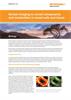
플라스틱 혈액백에 저장된 혈액의 연구에 사용된 inVia
캐나다 뱅쿠버 소재 브리티쉬 콜롬비아 대학교의 Michael Smith Laboratories에서 저장 적혈구(RBC) 유닛1,2에서의 생화학 변화와 기증자 간 변동성을 모니터링하기 위한 도구로 라만 분광기를 사용하는 방식을 선도하고 있습니다. Michael Blades와 Robin Turner 교수가 이끄는 연구진은 최근 Analyst에 이 연구 결과를 발표했습니다.
암 치료 도중 세포와 조직의 방사선 손상을 검출하기 위해 사용된 라만 분광기
캐나다 브리티쉬 콜롬비아 대학교의 Irving K Barber School of Arts and Sciences에서는 물리, 엔지니어링, 방사선 종양학 등 다양한 분야의 과학자들로 구성된 다학문 그룹이 연구에 참여하고 있습니다. 이 그룹은 암 치료에 사용되는 이온화 방사선에 의해 유발되는 세포와 조직 손상을 검출하고 이해하는 데 관심을 두고 있습니다.