현재 사용 중인 언어로는 이 페이지를 사용할 수 없습니다. Google Translate을 사용하여
자동 번역된 페이지
를 볼 수 있습니다. Renishaw에게는 이 서비스를 제공할 책임이 없으며 번역 결과를 저희가 확인하지도 않았습니다.
추가로 도움이 필요하시면
저희에게 연락해 주십시오.
Raman imaging of a worm
A non-invasive and label-free technique such as Raman spectroscopy, which can resolve a wide range of chemical details and simultaneously provide spatial information, is an ideal tool for studying model organisms.
11 October 2011
Nematode - model organismSimple multicellular organisms, such as nematode, zebrafish, and fruit flies, play a critical role in translational medicine. Their ease of breeding, rapid turnover rates, simple cellular organisation, and transparency make them ideal for understanding early developmental pathways, gene functions, and human diseases.
A non-invasive and label-free technique such as Raman spectroscopy, which can resolve a wide range of chemical details and simultaneously provide spatial information, is therefore an ideal tool for studying these organisms.
Nematode Caenorhabditis elegans is one of the key, and best, model organisms. Current biological assays for investigating biochemical changes in the nematodes post genetic modification are largely invasive and/or rely on the use of labels. These assays typically lack the capability to simultaneously quantify biochemical molecules and resolve spatial information.
Raman imaging was applied directly to unfixed Steinernema kraussei, a nematode species similar to C. elegans, to demonstrate the capabilities of Raman spectroscopy. Streamline Plus™ and StreamHR™ imaging were applied to the whole and selected areas of the same organism.
The Raman images provide valuable information on the biochemical species within the nematode and their spatial arrangement. By analysing the information provided by the Raman images and spectra, a multitude of phenotypic changes in the nematodes can be deduced without the need for labelling or dissection. Raman imaging therefore serves as an ideal method for studying chemo-structural changes in situ in micrometre scale model organisms.
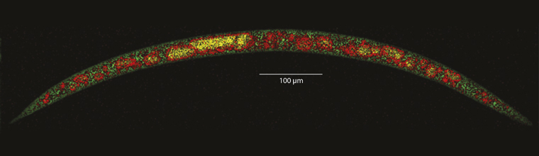
Above - A Raman image of the whole nematode based on the intensity variation of three Raman bands. Green: 1003 cm-1, ascribed to ring breathing mode of phenylalanine; yellow: 1247 cm-1 to 1282 cm-1, ascribed to amide III modes of protein; red: between 1640 cm-1 to 1677 cm-1, ascribed to amide I mode of protein and (=C-H) of unsaturated fatty acids. Phenylalanine is a typically ubiquitous constituent of biological species. Thus, the green region represents the main nematode cavity. The red regions represent protein and lipid-rich domains, and the yellow region represents protein-rich domains.
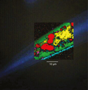 Right - Principal component anaylsis (PCA) performed on the tail region of the nematode unveiled information on the chemical species and their distribution, without labelling. In the figure, overlaid PCA images are superimposed on a white light micrograph. The green regions make up the bulk of the mapped area. According to the PCA loadings, they represent protein and lipid presence. The red regions represent lipid domains, where levels of protein presence are relatively low. The yellow domains are especially rich in protein of α-helix secondary structure and amino acids. The cyan regions correspond to the collagen rich region which correlates to the outer cuticle of the nematode. The magenta regions represent myoglobin presence.
Downloads
- Nematode - model organism [44kB]
-
 Nematode full worm image
[92kB]
Nematode full worm image
[92kB]
-
 Nematode cross-section worm image
[340kB]
Nematode cross-section worm image
[340kB]

All images and text copyright Renishaw.
News updates
Register for regular news updates from Renishaw.