현재 사용 중인 언어로는 이 페이지를 사용할 수 없습니다. Google Translate을 사용하여
자동 번역된 페이지
를 볼 수 있습니다. Renishaw에게는 이 서비스를 제공할 책임이 없으며 번역 결과를 저희가 확인하지도 않았습니다.
추가로 도움이 필요하시면
저희에게 연락해 주십시오.
Raman spectroscopy for oncology research (and drug delivery research)
March 2014
Raman microscopy is routinely used for characterisation and identification of material, but the need for this molecular imaging and analysis technique has become increasingly important in biology.
Renishaw Inc (USA) recently contributed to a webinar about Raman spectroscopy for oncology research (and drug delivery research). This proved very popular, with attendees learning about:
- a Raman technique used to identify cancer cells from healthy cells
- a Raman technique used to analyse lipid based drug discovery
- set up of an experiment using label-free detection (no fluorescent dyes, colorimetric stains or labelled antibodies needed)
The webinar is available to view on Lab Manager's website.
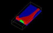
Image: Multiple component 3D Raman volume image of glioma cell. This shows the substrate (blue), cell (red) and nucleus (green). Renishaw thanks Dr Matthew Baker, University of Central Lancashire, for providing the cell sample.
News updates
Register for regular news updates from Renishaw